 |
Obstetrical
and Stethoscopes
|
In the pre-1900 era, most doctors knew how to
deliver a baby, but in practice it seems many were either delivered by lay
people or mid-wives. The instruments shown on this page illustrate the
more technical side of obstetrical delivery.
Early obstetrical forceps-
made of blue gun steel, c.1840; a later example from C.E. Kolbe, Phila. c.
1880; by Sharp and Smith, c. 1895; c.1929 by Sklar, chrome plated type.
| Forceps marked Tiemann & Co. Note the
adjustment screw in the distal of the handle. |
 |
| Quadruple dilator, plated, Weiss marked. |
 |
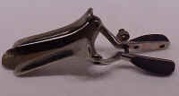
| Rare Hernstein marked tri-valve speculum with
crosschecked handles |
 |
| Rare unplated forceps with rich dark patina, marked
as made by Madera, (sp); (American made) Sixteen inches
long. |
 |
| Plated forceps made by C.W. Kolbe, Phila.
Sixteen inches long. |
 |
| RARE Small size forceps, marked Hernstein, New
York, composite handles, large intervals in the crosshatching.
Plated. Eleven inches long. |
 |
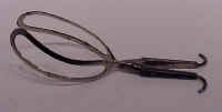
| RARE Small size forceps, marked Shepard &
Dudley. Tight crosshatching on the composite handles. Unplated. Nine
inches long. |
 |
| Speculum with wood handle, marked Shepard and
Dudley. Brass showing from cleaning plating. |
 |
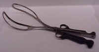
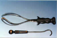
| RARE Bedford forceps, by Shepard and Dudley
c. 1875: note the finger holes on the larger standard size ebony handled forceps and
the very small forceps below with ebony handles. Bedford's forceps were introduced
in 1846 by Gunning S. Bedford (1806-1870). He was born in Baltimore and practiced in
New York. |
 |
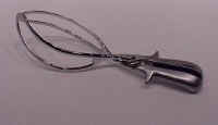
| Sklar forcep. All metal and from the 1930's.
|
 |
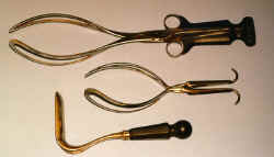
| Bedford forceps, by Shepard and Dudley
c. 1875: note the finger holes on the larger standard size ebony handled forceps and
the very small forceps below with ebony handles. The ebony handled speculum is also
part of this set. Note: Bedford's forceps were introduced in 1846 by Gunning S. Bedford
(1806-1870). He was born in Baltimore and practiced in New York.
|
 |
|
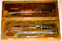
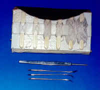
Cased English
obstetrical set, c. 1870 by Henry Lewis, London
Contains scalpel, delivery forceps, craniotomy forceps,
perforator, blunt hook and crochet. Some of the metal has been buffed to clean off
rust. A rare complete set in great condition.
|

 |
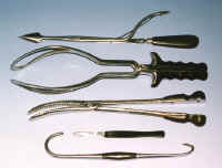 |
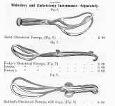
| Various obstetrical forceps from an 1860
Snowden and Brother catalog as typical of Civil War era instruments. |
 |
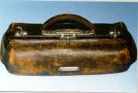
Historical note: Think about this.
It's 1880, there is a breech birth, the fetus is deceased, and the mother is in danger of
dying. What are the options? There is ether for an anesthetic, but antibiotics
and surgical knowledge don't exist to justify a c-section. The obstetric leather bag
above was taken to the home of the patient and gave the doctor a way out to save the
mother. Brutal times, but necessary.
Due to the presence of rickets in both Europe
and the states, it is estimated that about 14% of women at the time could not give natural
birth due to a smaller or malformed pelvis. Among populations without that
ailment, the problem was almost non-existent due to normal pelvis size.
|
Instruments
of destruction.
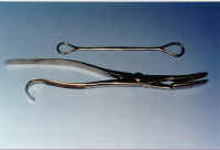
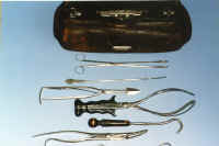
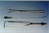
A
complete leather obstetrical case, c. 1880: by Sharp and Smith, included are ebony handled
delivery forceps, cranioclast forceps, cranial perforator with arrow head, blunt
hook with ebony handle, and other instruments for field delivery. Includes doctor
owner's name on letter and receipt. There are several other items in the case such
as hand soap, sulfa drugs, suture needles, etc.
Also, an episiotomy suture set
with silk sutures and needles found in the carrying bag. Please see the drug case
elsewhere on this site which was part of this set for the same doctor.
|
 |
 |
|
 |
 |
|
 |
 |
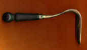
| Obstetrical retractor (speculum) by
Shepard and Dudley, c. 1875, plated, brass showing |
 |
|
Three way vaginal speculum with
ebony handles by A.L. Hernstein, N.Y., c. 1870's |
 |
|
Weiss mechanical uterine specula. Plated
brass, c. 1890's. |
 |
|
Tiemann & Co. vaginal speculum-
retracting and expanding types. Plated brass, c. 1890's |
 |
|
Pessaries: a set of hard
rubber uterine supports, c. 1880, G. Tiemann Co., NY |
 |
Historical note: Perhaps due to multiple
childbirths, the uterus would prolapse due to weakened or stretched ligaments and cause
pressure on the bladder. The pessary was used intra-vaginally to support the uterus
in a raised position.
Historical note: The
widespread existence of syphilis and gonococcal disease in the Western world during the
1800's could account for the necessity of dilation of the urethra because of scarring and
tissue damage from disease. The other reason...prostate gland swelling. As
you can imagine, if one had the "problem", relief was an urgent matter.
Most "medical" kits contained instruments like the above
"sounds" for solving the problem of urinary tract stricture by the process
of blunt, progressive dilation. These sounds were also used to locate kidney stones
in the bladder.
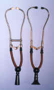
| Binaural stethoscopes:
Right - marked "Shepard and Dudley", c.
1870 with ebonized wood. This is the type which used an elastic band to constrict
the ear pieces. Elastic missing.
Left- c. 1876, with ivory ear pieces, German silver
tubes, ebonized wood bell, and silk wrapped India rubber tubing. Marked as
"Caswell Hazard & Co. - Ford" |
 |
|
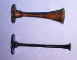
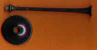
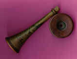
Monaural stethoscopes:
A 19th century 7" (longer and thinner of the two)
monaural ebonized wood stethoscope marked: Young, Edinb.
An early wood monaural stethoscope: in use
during the early 1800's. The bell at the top of the photo was placed against the
chest with the other end against the ear. The tube is hollow. This one
is made of a burl wood and the ear piece is detachable. |
 |
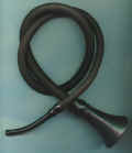
| Conversation tube (hearing aid) c. late
1800's, however, it appears to be very close to the monaural tubular stethoscopes in some
catalogs of the period. Unmarked.
|
 |
| An anesthetic vaporizer which was placed
over the nose and mouth where liquid ether would vaporize from the gauze when inserted in
the metal holder. The gauze was stretched back and forth over the insert. One
of the earliest anesthesia masks that could be easily sterilized with better control than
a simple handkerchief or mask. There was less CO2 contamination than a closed system
inhaler. Invented by S.H. Allis of Philadelphia in 1874. c. 1890 |
 |
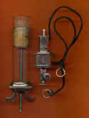
| A Heurteloup
artificial leech, c. ?. Mfg. unknown, but it is a part of the Japanese marked set
shown elsewhere on site. The exact items are shown in Edmonson's American
Surgical Instruments on page 53 and indicates its manufacture as Gemrig c.
1870. This is a real mystery as Gemrig was an American maker! |
 |
Historical note:
Bloodletting was practiced from the earliest periods of medical history. It was
believed that removing the "bad humors", and blood being a humor, from the
body would cure all sorts of maladies. If it worked for humans, then why not animals
too? They did bloodlet animals. George Washington was supposedly bled to
death by his doctors.
|
|