NEUROSURGICAL ANTIQUES

|
DOUGLAS ARBITTIER, M.D. In a previous article I discussed some historical notes about bloodletting through the ages and then went over some pointers about the artifacts of phlebotomy, illustrating some collectible bloodletting antiques. This article will briefly discuss the history of neurosurgery and illustrate some of the artifacts used in surgery on the head. Many individual items are illustrated as well as some neurosurgical sets. I’d like to point out that all of the individual items shown below are being offered for sale as a collection. The sets are not for sale. Please see the information at the end of the article if you are interested in purchasing this collection. The origins of neurosurgery can be traced to Neolithic times when it was presumably felt that boring a hole in the skull would release evil spirits or possibly cure other ailments. Of course, much of this is conjecture, but skulls of Neolithic men (and women) have been found that have clearly shown signs of brain surgery in life. Many ancient Peruvian skulls (over 4000 years old) have been found showing evidence of trepanning, where a depressed skull fracture was removed. A large number of these skulls have well-healed wounds, suggesting that people often survived the operation. This is remarkable, considering that the tools used were chipped stone knives and copper blades, and that the lack of antibiotics made infection very likely. Of course, a depressed skull fracture causing a subdural hematoma (bleeding on the brain) would have meant almost certain death, so trepanation was necessary to save lives, which it undeniably did on occasion. The ancient Greeks had "trepanons" or early bone drills which may have been used for boring into the skull. Much medical knowledge at that time was based on the four humors of the body (see my bloodletting article) and perhaps removing certain humors from the cranium was a basis for their practice. The earliest specialized skull saws used in England date to the 14th century. These were composed of a crescent-shaped blade with outer curve serrations. This design made it possible to cut a convex surface (eg. the skull) and manuscripts from Oxford, England attest to their use, sometimes successfully. Sir William Hey (1736-1819) spent years developing an improved skull saw in Leeds, England. His work came to fruition in 1803 when he finalized the saws that came to be known as Hey’s saws. Since that time no large general surgical or specialized neurosurgical set was complete without at least one of Hey's saws. These often had one or two straight or curved blades which were used to cut the cranium around a skull fracture until the dura, or brain covering, was exposed.
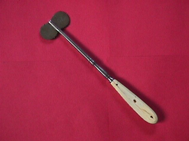
Figures 1 and 2 show some examples of Hey’s saws. Figure 1 shows two wooden handled instruments. The left has a hatched ebony handle impressed with the name Weiss and dates to ca. 1840. Note the slightly different curves to the two blades. The other Hey's saw is unusual. It has a mahogany handle (not commonly used) and two odd shaped blades. It has an unreadable signature but was made in Glasgow and dates to ca. 1850. Figure 2 shows a very rare and large (9.5 inches long while the others are 6.5 inches long) Hey's saw. It is heavier than the average Hey's saw and dates to the 1830s at the latest, but may possibly be somewhat earlier. While Hey's saws were most likely used to cut circular or rectangular holes in the skull a seemingly more efficient way was with trepans or trephines. The trepan is a frame or brace with drill bits that was probably introduced in the 16th century by Andrea de Croce of Venice. Both detachable perforators and circular saw bits were used with the frame or brace. Often there was a central pin in the circular bit to "get things started." This was a dangerous addition since the central pin had the possibility of penetrating the brain (a very bad idea) so the pin had to be removed.
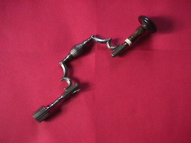
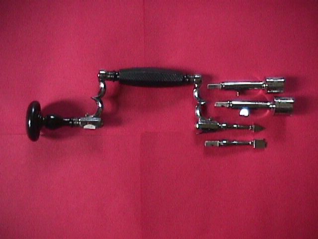
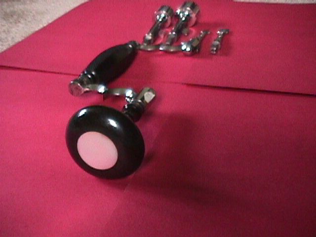
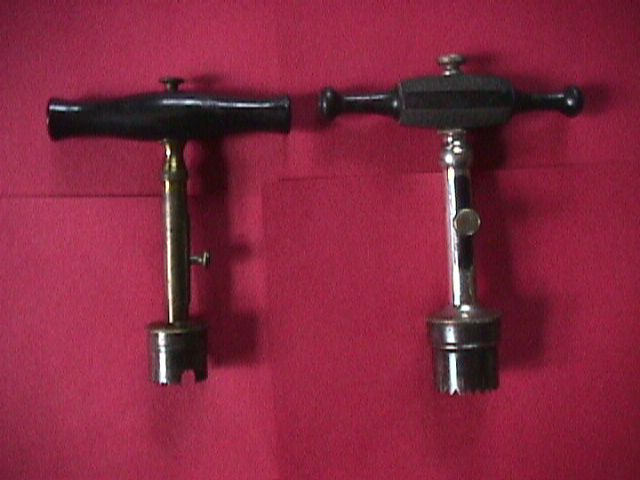
Figures 3 through 5 show two very rare trepan braces. Figure 3 is a rare brace with a horn handle and ivory ferrule. It is the classic shape for ca 1760-1780. The circular head may be a replacement, but certainly fits well via the spring release lever. Figures 4 and 5 show a German trepan with four interchangeable bits by Lutter of Germany ca 1870. The grip handle is hatched ebony and the head has a mother of pearl infill as the photo shows. This is an extraordinary decorative touch not often seen on similar trepans. This trepan is in mint unused condition which is most unusual considering its age. While the trepan brace was a sometimes cumbersome two handed instrument, the 16th century saw the development of the trephine which could be operated with one hand. Fabricius (1537-1619) first described the use of this T-shaped skull drill. Samuel Sharp improved on this by developing a central pin that was removable to prevent brain penetration. The trephine survived in one form or another well into the 20th century. The handles of these instruments were made of many materials and examples can be found in figures 6-10.
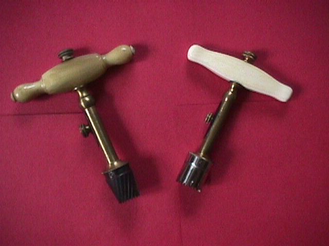
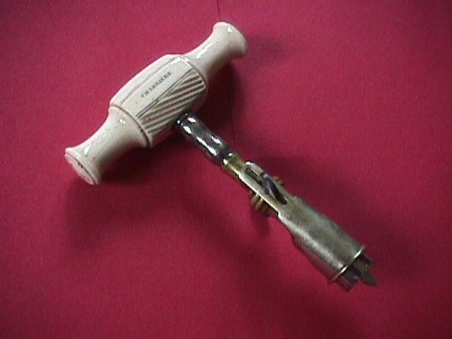
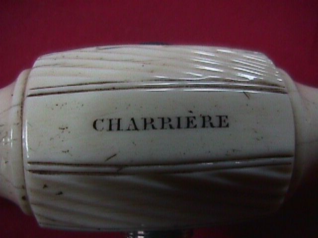
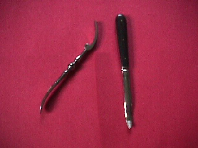
Figure 6 shows two ebony handled trephines, one with a smooth handle (ca 1830) and one with a hatched handle (ca 1860) for a better grip. Both have adjustable center pins. This is thought to be an invention of Rudtorffer of Vienna in 1817. ( Editor: Alex Peck has two references from the late 18th century that show adjustable pins.) The slits in the circular heads of these trephines were introduced by Benjamin Bell in 1801 as a way to disperse the inevitable bone dust created from drilling. Figure 7 shows two trephines with very decorative handles, dating to the mid 1800’s. One has a beautifully turned horn handle and one is a rare ivory handled trephine by Tiemann, a prolific and high quality New York maker. Figures 9 and 10 show perhaps the finest trephine ever produced. This is an exhibition standard nickel plated and brass trephine with a large and heavy ivory handle signed Charriere. Charriere was one of the premier French 19th century instrument makers who was active most of that century. The adjustable blade has a sliding collar to regulate depth which is controlled by two blued steel thumb screws. This instrument came from the Charriere family collection and is one of a kind which was presented at an international exhibition in 1867...truly a work of art. Figure 8 shows two all steel trephines, one signed Sharp and Smith, that demonstrate how the beauty of these instruments was lost after the age of sterilization, when ebony, ivory, and horn instruments fell by the wayside. Once a trepan or trephine was used to bore the hole in the skull an elevator was used to lift up the cut bone flap. Figure 11 shows two elevators, one a rare steel curved elevator c. 1770 and one with an ebony handle ca 1860.
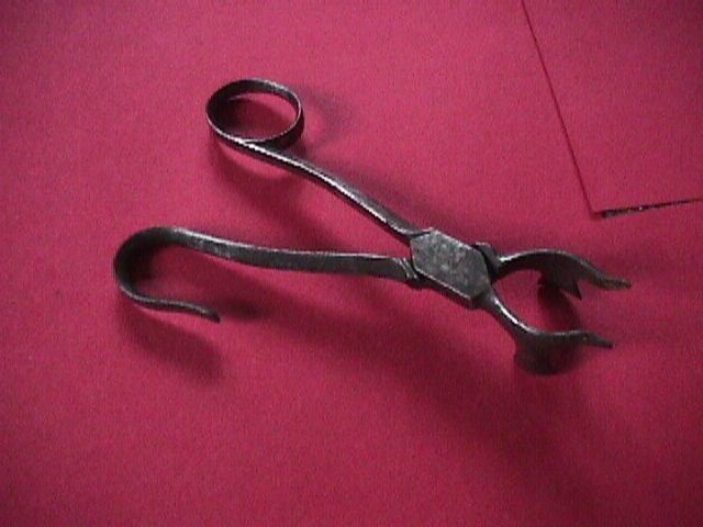
Samuel Sharp invented a special type of forceps in the early 1700’s that was used to lift up the circular piece of bone. One of these instruments is pictured in figure 12. The instrument was placed around the circular piece of bone to lift it off the brain. These forcep type instruments are extremely rare today. In order to smooth the edge of the circle cut from the skull, surgeons used file-like instruments called lenticulars. These had a smooth end with a sharp blade around it to depress the brain without causing soft tissue damage and still be able to file the bone edges. Frequently the circle of bone would fracture as it was removed and jagged edges were left which needed to be smoothed. Figure 13 shows two very rare examples of 18th century lenticulars, one with an ebony handle and one in ivory. Figure 14 shows an example from about 1840.
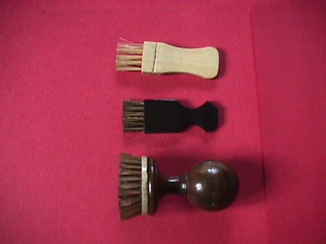
Drilling and cutting bone created bone dust which would have to be removed from the from the cut site as well as the drill and crowns. Early on the bone brush was invented to clear these particles from the instruments. Figure 15 shows three unusual bone brushes. The ebony and ivory handled examples are from the 19th century. The rounded one has an ivory dot on the top and is likely 18th century. It is made from two different woods. All are in excellent condition for their age.
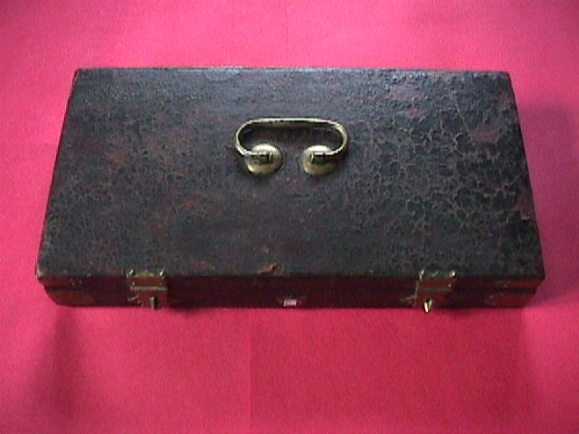
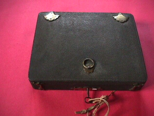
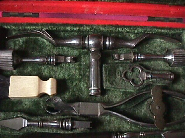
Perhaps the most remarkable surgical sets ever made were those specialized for neurosurgery. The following figures show some rare examples of these sets. Figures 16 and 17 are of a set c. 1780 made by Samson of Paris. It is complete and includes a trepan brace, drill bits, bone brush, lenticulars, elevators, etc.
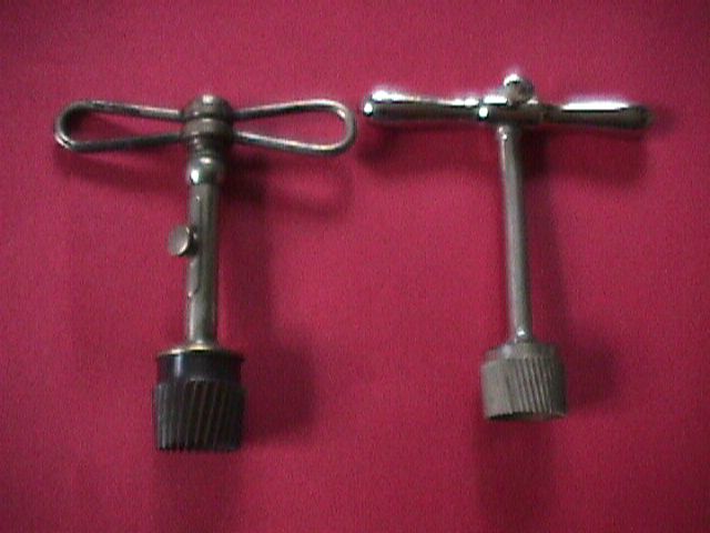
Figures 18-20 show a very rare cased set by Stanton, an English instrument maker. In this set is an example of Samuel Sharps gull wing type trephine which was mentioned earlier. This trephine has an elevator built into the T-handle. Edward Stanton was actively producing instruments in 1738, but there is no trace of his work in the literature after 1744.
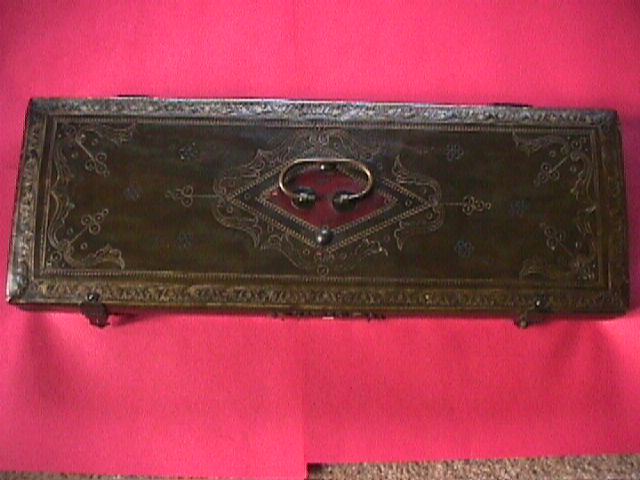
The greatest neurosurgical set I have ever seen is pictured in figures 21 and 22. This set may have been made by Grangeret of France and could date from the 18th century. It is very early and very fine. The gold tooled leather case and instruments are unused. The set includes multiple trepan saws, lenticulars, and elevators. This was certainly a decorative set made to be used on a discriminating clientele, perhaps royalty. The bone brush may be a replacement, though the one shown fits perfectly in its case space. Collecting neurosurgical antiques has become more difficult (as with all antiques) over the last decade. Where there was once a steady stream of such antiques coming to America it is now only a trickle. The dealers in medical antiques listed on this site are an excellent resource for this specialized area of medical antiques. They may be able to help one locate a special instrument or set. Prices of these items range from $50 for post sterilization trephines to many thousands of dollars for fine sets. Instruments in ivory are at a real premium. The above collection was put together over 16 years of careful and extensive searching. The pieces were collected with an eye toward excellent condition and rarity. Most of them fit this bill. The focus of my collecting has changed over the years (see below) and I have decided to sell all individual instruments listed above. Please email me if you want further details or have specific questions about individual instruments. References: Antique Medical Instruments by Elisabeth Bennion, Sotheby's
Publications, 1979. |